Mast Cell Activation Syndrome (MCAS) and the Microbiome
What is Mast Cell Activation Syndrome (MCAS)?
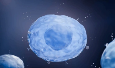
To understand MCAS, you first need to understand mast cells. Mast cells are a type of immune cell that guard your body against foreign objects. They are typically abundant in skin, near blood vessels and lymph vessels, nerves, lungs and intestines. When activated, they will release chemicals, also called mast cell mediators. The most well known mediator is histamine, but it is really important to note that mast cells have 390 known mediators.1
Mast cells are naturally activated in response to allergens and pathogens. In MCAS, mast cells are essentially much more easily activated. This leads to a chronic excess of mast cells mediators. Depending on what tissue is affected the most, this can present in a wide variety of different ways.
Symptoms may include2:
- Skin: hives, flushing, swelling
- Respiratory tract: wheezing, asthma, runny nose, shortness of breath
- Digestion: nausea, vomiting, diarrhea, abdominal pain, food reactions, heartburn
- Cardiovascular: low blood pressure, fainting, fast heart rate
- Other: joint pain, nerve pain, insomnia, brain fog, fatigue and more
When it comes to MCAS, individuals tend to have only a handful of symptoms, so it can look different in different people. Symptoms can wax and wane and symptoms can change over time. That is why it is hard to recognize in clinical practice. Diagnosing MCAS can be difficult due to lack of consensus on diagnostic criteria and challenges around testing (lab errors resulting in false negatives).
One reason people may have MCAS is genetics. The most well studied mutation is in the Kit gene. This gene encodes a receptor on the outside of mast cells that is involved in mast cell activation. Research has identified more than 50 different Kit mutations associated with different dysfunction in the mast cell.3,4 This results in problems with different mediators meaning symptoms will be different depending on the specific mutation. Testing for these mutations is not commercially available yet.
If genetics is the primary cause of MCAS, these individuals typically have a long history of illness and symptoms going back to their childhood. But what about the people who were healthy and then something happened and they acquired all these new symptoms that may be categorized as MCAS? Common presentations in these cases include onset after antibiotics, mold exposure, tick bite, or viral illness. For these individuals, MCAS may not be genetic, but essentially environmental — pathogens and toxins are triggering mast cells. For certain individuals, targeting the microbiome may be critical for resolving MCAS.
Mast Cells & the Gut
In the gut, mast cells act as sentinels. The gut is where the outside world interfaces with your immune system and your body. Mast cells are there to guard you. In the gut, mast cells have multiple roles. One role is to communicate with the nervous system in the gut —this can influence blood flow to the gut as well as gastrointestinal motility.5 This also impacts pain perception in the digestive tract.5 Mast cell mediators may also increase intestinal permeability and can activate other immune responses in the gut.5 So inappropriate mast cell activation could significantly impact the digestive tract. This can present as a wide variety of digestive symptoms, food reactions, and systemic inflammation.
How the excess mast cell mediators in the digestive tract affect the microbiome is not yet known. In conditions such as IBS where research has identified high numbers of colonic mast cells and more mast cell activation, we do see an altered microbiome.9 However, the alterations in the microbiome may not be due to the mast cells. In fact, the alterations in the microbiome may be the cause of the higher number of mast cells and more mast cell activations. Research in mice demonstrated that transplanting the IBS microbiome caused IBS in healthy mice9, suggesting that the microbiome may influence mast cells more than mast cells influence the microbiome.
Mast Cells & the Microbiome
Microbes can interact with mast cells through various mechanisms which impact mast cells maturation, activation, and function. One example is lipopolysaccharide (LPS), produced by gram negative bacteria such as Proteobacteria. In vitro studies show that LPS caused mature mast cells to increase expression of tryptase, chymase, IL-1beta and IL-6.6 Other in vitro studies indicate that LPS strongly activated mast cells and resulted in increased histamine.7
So the first microbiome consideration for individuals with MCAS is Proteobacteria overgrowth. This can happen in the small intestine or the large intestine. Small intestine bacterial overgrowth (SIBO) is common in MCAS.8 The exact relationship is unknown. One theory is that MCAS may cause SIBO by altering GI motility, but we also know that LPS from the overgrown bacteria in hydrogen SIBO are potent mast cell activators. Either way, clearing SIBO, if present, goes along way to improving both digestive and systemic symptoms in MCAS patients. Some individuals will experience resolution of MCAS symptoms once SIBO is clear.
Peptidoglycan is another bacterial component that can interact with mast cells. Peptidoglycan is the main component of bacterial cells walls found in gram positive bacteria. In vitro studies showed that peptidoglycan inhibits growth of mast cells.6 Other studies in the skin found that peptidoglycan (specifically from Staphylococcus aureus) caused mast cells to induce a Th1 immune response.10 Other studies (in vitro and animal models), have found different Lactobacillus species such as Lactobacillus paracasei to suppress degranulation of mast cells induced by IgE antibodies.11 This may be due to Lactobacillus peptidoglycan binding receptors on mast cells.
Research has also explored how Candida albicans interact with mast cells. In vitro studies showed that mast cells may be able to break down or kill Candida albicans.11 Interactions between Candida and mast cells also resulted in nitric oxide production, reactive oxygen species, and degranulation of mast cells.11 In human mast cells, pro-inflammatory and anti-inflammatory cytokines were released.11
Butyrate is a metabolite of a healthy microbiome that may have beneficial impacts on mast cells. Animal studies indicate that butyrate may inhibit mast cell degranulation and cytokine production.12 Other studies have found that butyrate also decreases mediators inside the mast cell and suppressed proliferation of mast cells.13 So butyrate may results in less mast cells, with less mediators that are more stable. But human studies are needed to explore how butyrate may help in MCAS.
While research has uncovered a lot about how mast cells and the microbiome interact, a whole lot more research is needed, particularly in humans, to better understand what this may mean for treatment of MCAS.
Some places to start may involve:
- Testing for SIBO and treating if positive
- Decreasing Proteobacteria in the large intestine if elevated
- Increasing butyrate producers in the large intestine if low
- Supplementing with specific probiotics that may stabilize mast cells such as Lacticaseibacillus casei14 , Lactiplantibacillus plantarum HD02 and MD15915, Lactobacillus rhamnosus JB-116, Lactobacillus rhamnosus GG (LGG) and Lc70516
Please be sure to talk to your doctor about what testing and treatment may be appropriate for you.
References
- Molderings GJ, Afrin LB. A survey of the currently known mast cell mediators with potential relevance for therapy of mast cell-induced symptoms. Naunyn Schmiedebergs Arch Pharmacol. 2023 Nov;396(11):2881-2891. doi: 10.1007/s00210-023-02545-y. Epub 2023 May 27. PMID: 37243761; PMCID: PMC10567897.
- Akin C. Mast cell activation syndromes. Journal of Allergy and Clinical Immunology. 2017;140(2):349-355. doi:10.1016/j.jaci.2017.06.007
- Molderings GJ, Kolck UW, Scheurlen C, Brüss M, Homann J, Von Kügelgen I. Multiple novel alterations in Kit tyrosine kinase in patients with gastrointestinally pronounced systemic mast cell activation disorder. Scand J Gastroenterol. 2007 Sep;42(9):1045-53. doi: 10.1080/00365520701245744. PMID: 17710669.
- Molderings GJ, Meis K, Kolck UW, Homann J, Frieling T. Comparative analysis of mutation of tyrosine kinase kit in mast cells from patients with systemic mast cell activation syndrome and healthy subjects. Immunogenetics. 2010 Dec;62(11-12):721-7. doi: 10.1007/s00251-010-0474-8. Epub 2010 Sep 14. PMID: 20838788.
- Intestinal mast cells in gut inflammation and motility disturbances - ScienceDirect. Accessed October 24, 2024. https://www.sciencedirect.com/
science/article/pii/ S0925443911000718 - Kirshenbaum AS, Swindle E, Kulka M, Wu Y, Metcalfe DD. Effect of lipopolysaccharide (LPS) and peptidoglycan (PGN) on human mast cell numbers, cytokine production, and protease composition. BMC Immunol. 2008 Aug 7;9:45. doi: 10.1186/1471-2172-9-45. PMID: 18687131; PMCID: PMC2527549.
- Wang Y, Sha H, Zhou L, Chen Y, Zhou Q, Dong H, Qian Y. The Mast Cell Is an Early Activator of Lipopolysaccharide-Induced Neuroinflammation and Blood-Brain Barrier Dysfunction in the Hippocampus. Mediators Inflamm. 2020 Feb 24;2020:8098439. doi: 10.1155/2020/8098439. PMID: 32184702; PMCID: PMC7060448.
- Weinstock, Leonard B. MD, FACG1; Brook, Jill MA2; Kaleem, Zahid MD3; Afrin, Lawrence MD4; Molderings, Gerhart MD5. 1194 Small Intestinal Bacterial Overgrowth Is Common in Mast Cell Activation Syndrome. The American Journal of Gastroenterology 114():p S671, October 2019. | DOI: 10.14309/01.ajg.0000594304.
61014.c5 - Shimbori C, De Palma G, Baerg L, Lu J, Verdu EF, Reed DE, Vanner S, Collins SM, Bercik P. Gut bacteria interact directly with colonic mast cells in a humanized mouse model of IBS. Gut Microbes. 2022 Jan-Dec;14(1):2105095. doi: 10.1080/19490976.2022.2105095. PMID: 35905313; PMCID: PMC9341375.
- 10.Matsui K, Kanai S, Ikuta M, Horikawa S. Mast Cells Stimulated with Peptidoglycan from Staphylococcus aureus Augment the Development of Th1 Cells. J Pharm Pharm Sci. 2018;21(1):296-304. doi: 10.18433/jpps29951. PMID: 30012242.
- 11.De Zuani, M., Dal Secco, C. and Frossi, B. (2018), Mast cells at the crossroads of microbiota and IBD. Eur. J. Immunol., 48: 1929-1937. https://doi.org/10.
1002/eji.201847504 - 12.Folkerts J, Redegeld F, Folkerts G, Blokhuis B, van den Berg MPM, de Bruijn MJW, van IJcken WFJ, Junt T, Tam SY, Galli SJ, Hendriks RW, Stadhouders R, Maurer M. Butyrate inhibits human mast cell activation via epigenetic regulation of FcεRI-mediated signaling. Allergy. 2020 Aug;75(8):1966-1978. doi: 10.1111/all.14254. Epub 2020 Apr 24. PMID: 32112426; PMCID: PMC7703657.
- 13.Folkerts J, Stadhouders R, Redegeld FA, Tam SY, Hendriks RW, Galli SJ, Maurer M. Effect of Dietary Fiber and Metabolites on Mast Cell Activation and Mast Cell-Associated Diseases. Front Immunol. 2018 May 29;9:1067. doi: 10.3389/fimmu.2018.01067. PMID: 29910798; PMCID: PMC5992428.
- 14.Iketani A, Takano M, Kasakura K, Iwatsuki M, Tsuji A, Matsuda K, Minegishi R, Hosono A, Nakanishi Y, Takahashi K. CCAAT/enhancer-binding protein α-dependent regulation of granule formation in mast cells by intestinal bacteria. Eur J Immunol. 2024 Oct;54(10):e2451094. doi: 10.1002/eji.202451094. Epub 2024 Jul 9. PMID: 38980255.
- 15.Kim AR, Jeon SG, Kim HR, Hong H, Yoon YW, Lee BM, Yoon CH, Choi SJ, Jang MH, Yang BG. Preventive and Therapeutic Effects of Lactiplantibacillus plantarum HD02 and MD159 through Mast Cell Degranulation Inhibition in Mouse Models of Atopic Dermatitis. Nutrients. 2024 Sep 6;16(17):3021. doi: 10.3390/nu16173021. PMID: 39275335; PMCID: PMC11396792.
- 16.Oksaharju A, Kankainen M, Kekkonen RA, Lindstedt KA, Kovanen PT, Korpela R, Miettinen M. Probiotic Lactobacillus rhamnosus downregulates FCER1 and HRH4 expression in human mast cells. World J Gastroenterol. 2011 Feb 14;17(6):750-9. doi: 10.3748/wjg.v17.i6.750. PMID: 21390145; PMCID: PMC3042653.
